
Prostate.Carcinoma.ai
More reports per hour.
Higher precision in every one.
Delivered in KSA by IMAGE Healthcare Middle East, subject to SFDA approval.
Learn the essentials about our software in 60 seconds.
Efficiency and Precision for Your Radiology
with Prostate.Carcinoma.ai
Take advantage of AI-based, automated MRI analysis in your daily radiology practice.
Prostate.Carcinoma.ai is the intelligent AI extension for prostate segmentation
in PACS systems and diagnostic DICOM viewers.
Routine is ours. The decision is yours.

Here are the benefits of using Prostate.Carcinoma.ai





Reduced time required:
Manually marking the prostate is a thing of the past. Automation frees up resources that can help address the shortage of healthcare professionals.
More accurate prostate volumetry:
Achieving more accurate calculation of prostate volume and PSA
density and improving urologists TRUS-guided biopsy with better
choice of treatment options due to consistent segmentation.
Less manual work:
Reducing radiologists‘ workload by automating manual processes can increase their job satisfaction.
Integration with your PACS or DICOM viewer:
Prostate.Carcinoma.ai is designed for integration into existing PACS and DICOM viewer environments, including IMAGE iQ-SYSTEM. Site-specific compatibility is confirmed before deployment.

Alexander Cornelius, M.D., Deputy Director of the Institute and Chief Radiologist, Head of Urogenital Radiology and MRI, Aarau Cantonal Hospital, Switzerland
“The biggest advantage is that we currently measure the prostate in two dimensions with very low accuracy and then interpolate the prostate volume from those measurements. With the FUSE-AI algorithm, the entire prostate is segmented, enabling a precise determination of its volume.
This is significantly faster than our previous method and, at the same time, much more accurate. This is particularly relevant because PSA density is calculated based on prostate volume. PSA density is an important risk factor in assessing the prostate for prostate cancer.”

Prof. Dr. Thorsten Persigehl, Head of the Department of Oncological Imaging, University Hospital of Cologne, Germany
"Prostate.Carcinoma.ai significantly reduces the radiologist's workload through automatic segmentation and volumetric analysis of the prostate."
From MRI Images to Precise Segmentation
In 4 steps to Prostate.Carcinoma.ai
-
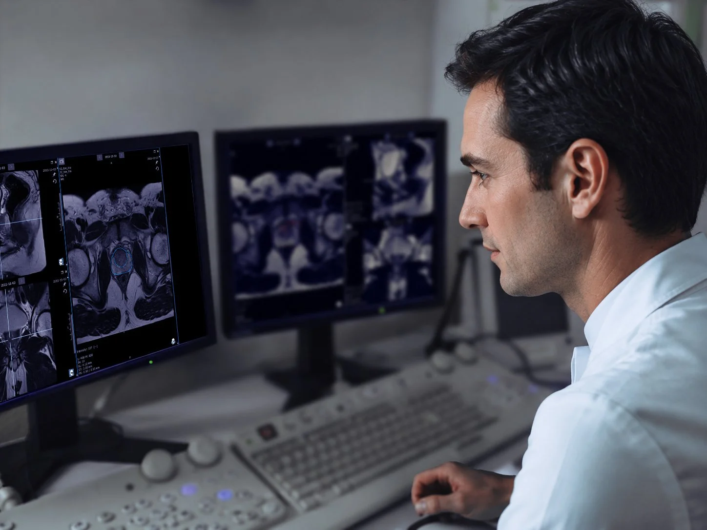
Prostate.Carcinoma.ai is an AI-powered software tool for the automatic segmentation of the prostate in MRI images. It assists radiologists with time-consuming manual tasks and can be integrated directly into PACS and diagnostic DICOM viewers.
-
The software automatically generates segmentations of the entire prostate as well as the peripheral and transitional zones. This eliminates a large portion of the manual contouring and zoning.
-
Automation reduces manual and error-prone steps, therebysaving time and reducing the workload in the radiology workflow. At the same time, it provides a consistent foundation for further interpretation.
-
By fully segmenting the prostate, the software enables amore accurate volume determination than a two-dimensional measurement followed by volume interpolation. Among other things, this provides a better basis for calculating PSA density.
-
Prostate.Carcinoma.ai is designed as a containerised software module and can be integrated into existing PACS and DICOM viewer environments, including IMAGE iQ-SYSTEM. The integration uses standard DICOM workflows. Site-specific compatibility and deployment requirements are reviewed before installation.
-
Prostate.Carcinoma.ai is medical device software certified under the European Medical Device Regulation, MDR (EU) 2017/745. FUSE-AI also operates a quality management system certified according to ISO 13485.
Prostate.Carcinoma.ai is not yet registered with the Saudi Food and Drug Authority and does not currently have Saudi market authorisation. The regulatory pathway for Saudi Arabia is being prepared together with our local partner.
-
The software was developed and validated in close cooperation with the Department of Radiology at Kantonsspital Aarau in Switzerland.
A local pilot or evaluation in Saudi Arabia may be considered together with an interested healthcare institution. Its scope and timing are subject to regulatory approval and the applicable contractual, technical and data protection requirements.
-
The standard deployment is on-premises, within the hospital’s IT infrastructure. Medical image files received by the software are stored only temporarily within the Docker container and are deleted after processing. DICOM data is anonymized before temporary storage.
If the software is deployed on servers in Saudi Arabia, all data processing remains within the hospital network and the Kingdom of Saudi Arabia.
-
The standard deployment is a Docker-based local installation on a server provided by the hospital or in its data center. Requirements include a Docker-compatible server with at least 32 GB of available RAM, appropriate storage access, a network connection, and the necessary DICOM connections and firewall settings.
The final server configuration depends on the expected volume of examinations, the clinical workflow, and PACS integration. It is determined prior to deployment as part of a technical coordination process. -
FUSE-AI and IMAGE Healthcare Middle East will coordinate onboarding, technical integration, user training and ongoing support. IMAGE Healthcare Middle East is intended to serve as the local first point of contact, while product-specific and advanced technical matters will be handled together with FUSE-AI. The detailed responsibilities and support arrangements will be agreed before deployment.
-
Prostate.Carcinoma.ai is offered under a licence model with contract options of one, three or five years. A pilot or evaluation may be considered before a commercial deployment, subject to regulatory approval and the technical, contractual and organisational requirements of the healthcare institution. The commercial conditions and exact licence scope are confirmed individually.
-
Prostate.Carcinoma.ai processes DICOM-compliant prostate MRI images and is designed for integration with PACS systems and diagnostic DICOM viewers, including IMAGE iQ-SYSTEM. Compatibility depends on the required imaging parameters and the supported DICOM workflow. Site-specific compatibility is technically verified before deployment.
FAQ

Book a demo and we'll show you how you can use AI to make your prostate MRI segmentation more efficient.
Please contact us!
We look forward to hearing from you—whether you have a question, would like us to call you back, or would like to schedule an appointment.
Your Contacts for the Middle East
Our team is happy to assist you with any questions you may have about Prostate.Carcinoma.ai, to provide a personalized product demonstration, and to offer technical and regulatory information.
We look forward to hearing from you!
Your contacts:

Mohamed Abouelkhiar
Managing Director
IMAGE Healthcare Middle East LLC

Ing. Majd Alismaeil
QM & RA Manager
QMR
Middle East Contact
FUSE-AI



FUSE-AI GmbH
Großer Burstah 46/48
D-20457 Hamburg
Contact
info@fuse-ai.de
+49/40/450318-0
www.fuse-ai.de

Subscribe to our newsletter!